On this page I will present you what things we, physiotherapist, need to do before starting any program of rehabilitation of shoulder injury or condition. Before we start complex evaluation of shoulder joint injuries and conditions, let us go back to basics and check out how one assessment form looks:


After comprehensible evaluation let us check the key findings in the history and physical examination:

Now we can start with shoulder range of motion (ROM):


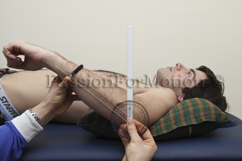
The alignment of the goniometer at the end of the ROM in
the shoulder complex extension. The examiner's hand that formerly stabilized
the subject's trunk now positions the goniometer.


Anterior and posterior drawer test can be done in supined position also. The arm of the patient is abducted and placed over the edge of plinth. The examiner stabilizes the scapula with one arm whilst the other grasps the humeral head and translates it in an anteromedial direction on the glenoid. Unilateral increases in humeral head translation of the symptomatic shoulder anterior GH instability. Posterior instability test is similar to the anterior draw test and the humeral head is translated in a posterolateral direction. A positive result is a unilateral increase in humeral head posterior translation on the glenoid. Diagnosis: Anterior or posterior GH instability.


This is an examination to dertemine the extent and/or presence of inferior instability of the glenohumeral joint. This test can be administered with the patient either standing or seated with their arm relaxed at their side. The examiner palpates the shoulder by placing thumb and fingers on the anterior and posterior aspects of the humeral head. The examiner grasps the patient's elbow with their other hand and applies a downward distraction force. A positive test will result in a sulcus being formed between the acromion and the humeral head as the humeral head moves inferiorly while the force is being applied.


The load and shift test as described by Hawkins is a modification of the anteriorand posterior drawer tests of Gerber and Ganz. It has been considered the gold standard for assessment of anterior and posterior instability.
The examiner creates a loading force to relocate the humeral head centrally in the glenoid. In this 'loaded position' directional stresses are applied. The examiner places one hand over the shoulder and scapula to stabilise the shoulder girdle and uses the other hand to grasp the humeral head. The humerus is loaded into the glenoid and then translated anteriorally and posteriorally. As the stress applied is increased the humeral head may be felt to ride up the glenoid rim. This test not only assesses the amount of translation but also provides an idea of the adequacy of the glenoid lip. It is critically important to compare the two shoulders to appreciate similarities or differences in translation.
The test is then repeated in supine. For this position the arm is grasped
and positioned in about 20° of abduction and forward flexion. The humeral head is again loaded then posterior and anterior stresses are applied. Although translation is assessed initially in the neutral position with the arm by the side it is important to assess translations in other positions as well. For example by progressively externally rotating the arm in the normal shoulder in abduction one should appreciate less translation anteriorly as the inferior glenohumeral ligament (GHL) becomes taut and acts as a restraint. Similarly by internally rotating the armposterior translation is diminished with an intact posterior capsular structure.
1. Supraspinatus tear test – Jobe’s Test


 The Infraspinatus muscle is one of the four rotator cuff muscles crossing the shoulder joint and is commonly injured. It is the main external rotator of the shoulder joint.

 Subscapularis is one of the four rotator cuff muscles which cross the shoulder joint. The muscle also acts to hold the head of the humerus in position and prevents it moving forwards. It is shown here with a couple of ribs cut away at the front.
Teres Major
 Teres major is only functional when theRhomboids fix the scapula. This muscle mainly helps Latissimus dorsi.
1. Empty can test
The Empty Can Test (ECT) was originally described by Jobe and Moynes to test integrity of the supraspinatus tendon. Kelly later proposed the Full Can Test (FCT) as an alternative as though EMG activity in the supraspinatus was similar in both positions the FCT was less provocative. It was therefore less likely to result in muscle weakness due to pain provocation.
Test:
The patient is tested at 90° elevation in the scapula plane and full internal rotation (empty can) or 45°external rotation (full can). Patient resists downward pressure exerted by examiner at patients elbow or wrist.
Positive :
FCT
Pain
Muscle weakness Pain/Muscle Weakness/Both
ECT
Pain
Muscle weakness Pain/Muscle Weakness/Both
Pain located to subacromial region and/or weakness.
In Itoi's study the two tests were performed in 143 shoulders of 136 consecutive patients. The tests were considered positive when there was pain, muscle weakness or both. Shoulders were then examined by high resolution MRI with 95% accuracy for full thickness tears of the supraspinatus tendon. There were 35 shoulders with full thickness tear of the supraspinatus tendon. The accuracy of the tests was the greatest when muscle weakness was interpreted as indicating a tomsupraspinatus tendon in both the full can test (75% accurate) and the empty can test (70%) accurate).
Itoi suggests that as the empty can position is more likely to be pain provoking, the full can test may be more beneficial in the clinical setting. Kelly, Kadrmas & Speer (1996) showed the best position for maximal isolation of the supraspinatus muscle was best achieved with the test position of elevation at 90° of scapular elevation and +45° (external rotation) of humeral rotation ('full can'). Howver, Boettcher, Ginn & Carruthers (2009) demonstrated on EMG studies that the supraspinatus is not sufficiently isolated in the empty can postion of abduction, calling into question the validity of this test for the diagnosis of suprapinatus patholgy. Suspect: impingement, tendonitis, supraspinatus tear
2. Neer’s test
Dr Neer developed this test based on his observations during shoulder surgery. He reported that the critical area for degenerative tendonitis and tendon ruptures was focused on the supraspinatus tendon and at times involved the anteriorinfraspinatus and occasionally the long head of biceps. Elevation of the arm in external or internal rotation causes critical areas to pass under the coraco-acromialligament or anterior acromion.
Test :
The examiner performs maximal passive abduction in the scapula plane, with internal rotation, whilst stabilising the scapula.
Positive :
Pain located to the sub-acromial space or anterior edge of acromion
False positive
Internal impingement Macdonald et al (2000)
- Bankart 25%
- SLAP 46.1 %
Research
Valadie' s paper demonstrated soft tissue contact with the medial acromion and contact between the articular surface of the rotator cuff and anterior- superiorglenoid rim.
Suspect: Subacromial impingement, rotator cuff pathology, biceps tendon.
3. Hawkins test
Originally described in the 1980's the Hawkins and Kennedy test was interpreted as indicative of impingement between the greater tuberosity of thehumerus against the coraco- humeral ligament, trapping all those structures which intervene. It has been reported as less reliable than the Neer impingement test.
Test:
The patient is examined in sitting with their arm at 90° and their elbow flexed to 90°, supported by the examiner to ensure maximal relaxation. The examiner then stabilises proximal to the elbow with their outside hand and with the other holds just proximal to the patient's wrist. They then quickly move the arm into internal rotation.
Positive
Pain located to the sub-acromial space
False Positive
- Internal impingement Macdonald et al (2000)
- Bankart 25%
- SLAP 46.1 %
Research
Valadie et al (cadaver study) describe consistent contact between the soft tissues and the coraco-acromialligament and between the articular surface of the rotator cuff and the anterior superior glenoid during the Hawkin' s and Kennedy test. Edelson and Teitz (2000) examined a large number of skeletal specimens and reported contact between the lesser tuberosity and antero- superior glenoid in the Hawkin' s and Kennedy test position. Roberts et al(2002) used MRI to identify and measure the changes in anatomic structures in the subacromial space as the arm was moved from complete rest to 160° of forward flexion during the Hawkin's and Neer impingement manoeuvres. The rotator cuff insertion appeared to be in closest proximity to the anteroinferioracromion not at full elevation (Neer sign position) but at 90° offlexion (Hawkin's sign position). Pain with this maneuver suggests subacromial impingement or rotator cuff tendonitis.
Suspect: subacromial impingement - rotator cuff tendonitis.
4. Subcoracoid impingement
Coracoid Impingement Sign - performed with the patient standing with the shoulder abducted 90 degrees with horizontal adduction in the coronal plane and maximally internally rotated to bring the lesser tuberosity in contact with the coracoid (the tennis "follow through" position).
Suspect: Subscapularis muscle - Subcoracoid impingement
5. Speed’s test (biceps b.)
The patient’s supinated arm is held at 90 degrees elbow flexion and then flexed forwards against resistance. Pain felt in the bicipital groove indicates biceps tendon pathology.
6. Belly press – subscapularis sign
Belly Press / Napoleon Sign - if patient cannot fully internally rotate and push on their belly, elbow will drop backwards if positive. The examiner pushes against the patient's elbows. (Laurent La Fosse)
7. Gerber’s lift off test (Gerber and Krushell 1991)
The dorsum of the patient’s hand is positioned at the level of the midlumbar spine. The subject is then asked to lift the dorsum of the hand off the back as far as possible, by internally rotating the shoulder. The test is considered positive for subscapularis dysfunction if the subject cannot lift the hand off of the back or if the subject performed the lifting manoeuver with elbow or shoulder extension. The test can be repeated whereby the patient is asked to try and push the examiner’s hand away from “hand behind back position”. A positive test is inability with or without pain.
8. O'Brien test (O'Brien et al. 1998)
The patient’s shoulder is held in 90 degrees of forward flexion, 30–45 degrees of horizontal adduction and maximal internal rotation. The examiner exerts a downward force distal to the patient’s elbow which the patient tries to resist. The patient is asked to identify, if produced, the location of the pain. The test is repeated in the same position except that this time the humerus is externally rotated and the forearm supinated, so the palm faces up. Once again, a downward force is applied by the examiner, which the patient actively resists, and the patient is asked to identify the location of any pain provoked. The test is considered positive if pain produced during the first part of the test is abolished with the second part of the test. For indication of a SLAP tear the pain is located over the anterior aspect of the shoulder, and for AC joint pathology, the pain must be located over the AC joint.
9. Anterior/posterior AC shear test (Davies et al. 1981)
With the patient sitting, the examiner cups the heels of both hands, one over the midpoint of the clavicle, anteriorly, and one over the spine of the scapula, posteriorly. With a compressive action both hands are squeezed towards each other. Several repetitions are applied with note being taken of the amount of movement compared with the opposite shoulder. Pain is also considered. A positive test is when the patient complains of superiorly located pain unilaterally.
10. Posterior Capsule Tightness
Measuring PC tightness: The subject is side lying, approximately half the distance of the length of the humerus away from the edge of the table. The non testing arm is placed under the head. The spine should be as close to neutral as possible. The medial epicondyle of the humerus is marked with a skin pencil. The tester stands facing the patient and grasps the limb just distal to the epicondyle. The humerus is brought to 90 degrees of abduction with no rotation of the humerus. The scapula is grasped and retracted at the lateral boarder. While the position of the scapula is maintained the humerus is slowly lowered into a horizontal adduction position. The humerus is lowered until motion has stopped or there is humeral rotation. When the motion is finished, a measurement is taken from the table to the mark on the elbow. This was shown to have excellent reproducibility because the scapular retraction allows for a reliable starting position every time. The researchers have found that for every 4 degrees f internal rotation lost, posterior capsule tightness will increase by 1cm using the method described.


After comprehensible evaluation let us check the key findings in the history and physical examination:

Now we can start with shoulder range of motion (ROM):
Shoulder
ROM
Lateral
rotation 90°
Medial
rotation 90°
Abduction
180°
Extension
55°
Flexion
180°

The end of the ROM of glenohumeral flexion. The examiner
stabilizes the lateral border of the scapula with his hand. The examiner is
able to determine that the end of the ROM has been reached because any attempt
to move the extremity into additional flexion causes the lateral border of the
scapula to move anteriorly and laterally.

The end of the ROM of shoulder complex flexion. The
examiner stabilizes the subject's trunk and ribs with her hand. The examiner is
able to determine that the end of the ROM has been reached because any attempt
to move the extremity into additional flexion causes extension of the spine and
movement of the ribs.

The alignment of the goniometer at the beginning of the
ROM of glenohumeral and shoulder complex flexion.

The alignment of the goniometer at the end of the ROM of
glenohumeral flexion. THe examiner's hand supports the subject's extremity and
maintains the goniometer's distal arm in correct alignment over the lateral
epicondyle. The examiner's other hand releases its stabilization and aligns the
goniometer's proximal arm with the lateral midline of the thorax.

The alignment of the goniometer at the end of the ROM of
shoulder complex flexion. More ROM is notet during shoulder complex flexion
than in glenohumeral flexion.

The end of the ROM of shoulder complex extension. The
examiner stabilizes the subject's trunk and ribs with his hand. The examiner is
able to determine that the end of the ROM has bees reached because any attempt
to move the extremity into additional extension causes flexion and rotation of
the spine.

The alignment of the goniometer at the beginin of the ROM
of glenohumeral and shoulder complex extension.

The alignment of the goniometer at the end of the ROM in
glenohumeral extension. THe examiner's left hand supports the subject's
extremity and holds the distal arm of the goniometer in correct alignment over
the lateral epicondyle of the humerus.


The end of the ROM of glenohumeral abduction. The
examiner stabilizes the lateral border of the scapula with his hand to detect
upward rotation of the scapula. alternatively , the examiner may stabilize the
acromion and coracoid processes of the scapula to detect elevation of the
scapula.

The end of the ROM of the shoulder complex abduction. The examiner stabilizes the subject's trunk and ribs with his hand to detect lateral flexion of the spine and movement of the ribs.

The alignment of the goniometer at the beginning of the
ROM in glenohumeral and shoulder complex abduction.

The alignment of the goniometer at the end of the ROM in
glenohumeral abduction. The examining table of the examiner's hand can support
the subject's extremity and align the goniometer's distal arm with the humerus.
The examiner's other hand has released its stabilization of the scapula and is
holding the proximal arm of the goniometer parallel to the sternum.

The alignment of the goniometer at the end of the ROM in the shoulder complex abduction. Note that the humerus is laterally rotated and the medial epicondyle is a helpful anatomical landmark for aligning the distal arm of the goniometer.

The end of the ROM of medial (internal) rotation of the
shoulder complex. The examiner stabilizes the distal end of the humerus to maintain
the shoulder in 90 degrees of abduction and the elbow in 90 degrees of flexion
during the motion. Resistance is noted at the end of medial rotation of the
shoulder complex because attempts to move the extremity into further motion
cause the spine to flex or rotate. The clavicle and scapula are allowed to move
as they participate in the shoulder complex motions.

The alignment of the goniometer at the beginning of
medial rotation ROM of the glenohumeral joint and shoulder complex.
The alignment of the goniometer at the end of the medial
rotation ROM of the glenohumeral joint. The examiner uses one hand to support
the subject's forearm and the distal arm of the goniometer. The examiner's
other hand holds the body and the proximal arm of the goniometer.

The alignment of the goniometer at the end of the medial
rotation ROM of the shoulder complex.

The end of lateral rotation ROM of the glenohumeral
joint. The examiner's hand stabilizes the spine of the scapula. The end of the
ROM in lateral rotation is reached when additional motion causes the scapula to
posteriorly tilt or retract and push against the examiner's hand.

The end of lateral rotation ROM of the shoulder complex
The examiner stabilizes the distal humerus to prevent shoulder abduction beyond
90 degrees. The elbow is maintained in 90 degrees of flexion during the motion.

The alignment of the goniometer at the beginning of
lateral rotation ROM of the glenohumeral joint and shoulder complex.

The alignment of the goniometer at the end of lateral
rotation ROM of the glenohumeral joint. The examiner's hand supports the
subjects forearm and the distal arm of the goniometer. The examiner's other
hand holds the body and proximal arm of the goniometer. The placement of the
examiner's hand would be reversed if the subject's right shoulder were being
tested.

The alignment of the goniometer at the end of lateral
rotation ROM of the shoulder complex.
Shoulder
Stability tests
1. Apprehension Test

Apprehension Test is performed in supine position with the humerus in 90 degrees of abduction, 90 degrees of elbow flexion and external rotation of the shoulder. The examiner exerts gentle pressure into progressive external rotation. A positive test is when the patient feels a sensation of impending dislocation. Diagnosis: Anterior GH instability
2. Relocation Test
2. Relocation Test

With the patient supine the arm is taken into abduction and external rotation. The test can be augmented by pushing the humeral head anteriorly from behind. The relocation test is performed by pushing posteriorly on the upper part of the humerus. The relocation test is positive if the apprehension or pain is relieved. Diagnosis: Anterior GH instability
3. Anterior and posterior stability testing – Drawer testing
 |
| Starting position |
 |
| Anterior Drawer |
 |
| Posterior Drawer |
Anterior and posterior drawer test can be done in supined position also. The arm of the patient is abducted and placed over the edge of plinth. The examiner stabilizes the scapula with one arm whilst the other grasps the humeral head and translates it in an anteromedial direction on the glenoid. Unilateral increases in humeral head translation of the symptomatic shoulder anterior GH instability. Posterior instability test is similar to the anterior draw test and the humeral head is translated in a posterolateral direction. A positive result is a unilateral increase in humeral head posterior translation on the glenoid. Diagnosis: Anterior or posterior GH instability.
4. Inferior stability testing - Sulcus sign


This is an examination to dertemine the extent and/or presence of inferior instability of the glenohumeral joint. This test can be administered with the patient either standing or seated with their arm relaxed at their side. The examiner palpates the shoulder by placing thumb and fingers on the anterior and posterior aspects of the humeral head. The examiner grasps the patient's elbow with their other hand and applies a downward distraction force. A positive test will result in a sulcus being formed between the acromion and the humeral head as the humeral head moves inferiorly while the force is being applied.
5. Load and Shift Test


The examiner creates a loading force to relocate the humeral head centrally in the glenoid. In this 'loaded position' directional stresses are applied. The examiner places one hand over the shoulder and scapula to stabilise the shoulder girdle and uses the other hand to grasp the humeral head. The humerus is loaded into the glenoid and then translated anteriorally and posteriorally. As the stress applied is increased the humeral head may be felt to ride up the glenoid rim. This test not only assesses the amount of translation but also provides an idea of the adequacy of the glenoid lip. It is critically important to compare the two shoulders to appreciate similarities or differences in translation.
The test is then repeated in supine. For this position the arm is grasped
and positioned in about 20° of abduction and forward flexion. The humeral head is again loaded then posterior and anterior stresses are applied. Although translation is assessed initially in the neutral position with the arm by the side it is important to assess translations in other positions as well. For example by progressively externally rotating the arm in the normal shoulder in abduction one should appreciate less translation anteriorly as the inferior glenohumeral ligament (GHL) becomes taut and acts as a restraint. Similarly by internally rotating the armposterior translation is diminished with an intact posterior capsular structure.
Special shoulder tests
1. Supraspinatus tear test – Jobe’s Test

The Supraspinatus muscle is one of the four muscles which make up the rotator cuff. Its main function is to stabilise the upper arm by holding the head of the humerus in position. It is important in throwing motions to control any forward motion of the head of humerus.
Origin: Supraspinous fossa
Insertion: Greater tuberosity of the humerus
Actions: Abduction, Stabilisation of the humerus
Innervation: Suprascapular nerve
Daily uses: Holding shopping bags away from the body
2. External rotation lag sign
The patient is seated. The elbow is passively flexed to 90 degrees and the shoulder is held at 20 degrees elevation in the scapular plane in a position of near maximum external rotation (i.e. maximum
external rotation minus five degrees to avoid elastic recoil). The examiner supports the elbow and holds the arm in external rotation at the wrist. The patient is asked to hold the position while the examiner supports the elbow but releases the hold at the wrist. The degree of movement is estimated and is referred to as the “lag” (i.e. the difference between active and passive ROM).
 |
| Starting position |
 |
| Negative |
 |
| Positive |
 The Infraspinatus muscle is one of the four rotator cuff muscles crossing the shoulder joint and is commonly injured. It is the main external rotator of the shoulder joint.
Origin: Posterior surface of the scapula (below the spine of the scapula)
Insertion: Greater tuberosity on the humerus
Actions: Shoulder abduction, External rotation
Innervation: Suprascapular nerve
Daily uses: Brushing hair
Teres Minor
Teres Minor is one of the four rotator cuff muscles surrounding the shoulder. Its main action, along with Infraspinatus is to externally rotate the shoulder joint. There are two Teres muscles, the other being Teres Major.
Origin: Mid section of the lateral border of the scapula
Insertion: Greater tuberosity on the humerus
Actions: External rotation, Shoulder adduction
Innervation: Axillary nerve
Daily uses: Brushing hair
3. Internal rotation lag sign test
The patient is asked to position his hand behind his back so that the dorsum of the hand is on the lumbar region. The examiner passively lifts the hand away from the lumbar region, whilst maintaining glenohumeral internal rotation. The patient is then asked to voluntarily maintain this position with only elbow support from the examiner. A positive result is if the hand falls back towards the spine, indicating a lesion of the subscapularis. The magnitude of the fall back can be recorded to the nearest 5 degrees.
The patient is asked to position his hand behind his back so that the dorsum of the hand is on the lumbar region. The examiner passively lifts the hand away from the lumbar region, whilst maintaining glenohumeral internal rotation. The patient is then asked to voluntarily maintain this position with only elbow support from the examiner. A positive result is if the hand falls back towards the spine, indicating a lesion of the subscapularis. The magnitude of the fall back can be recorded to the nearest 5 degrees.
 |
| Starting position |
 |
| Negative |
 |
| Positive |
Subscapularis
 Subscapularis is one of the four rotator cuff muscles which cross the shoulder joint. The muscle also acts to hold the head of the humerus in position and prevents it moving forwards. It is shown here with a couple of ribs cut away at the front.
Origin: Anterior (costal) surface of the scapula
Insertion: Lesser tuberosity of the humerus
Actions: Internal rotation, Adduction
Innervation: Upper and lower subscapular nerves
Daily uses: Tucking the back of your shirt into your trousers
Teres Major
 Teres major is only functional when theRhomboids fix the scapula. This muscle mainly helps Latissimus dorsi.
Origin: lower 1/3 of the lateral border of the scapula
Insertion: Intertubercular goove (between the greater and lesser tubercles) of the humerus
Actions: Adduction, Internal rotation, Extension
Innervation: Lower subscapular nerve
Daily uses: Tucking the back of your shirt into your trousers
Specific shoulder tests
1. Empty can test
The Empty Can Test (ECT) was originally described by Jobe and Moynes to test integrity of the supraspinatus tendon. Kelly later proposed the Full Can Test (FCT) as an alternative as though EMG activity in the supraspinatus was similar in both positions the FCT was less provocative. It was therefore less likely to result in muscle weakness due to pain provocation.
Test:
The patient is tested at 90° elevation in the scapula plane and full internal rotation (empty can) or 45°external rotation (full can). Patient resists downward pressure exerted by examiner at patients elbow or wrist.
Positive :
FCT
Pain
Muscle weakness Pain/Muscle Weakness/Both
ECT
Pain
Muscle weakness Pain/Muscle Weakness/Both
Pain located to subacromial region and/or weakness.
In Itoi's study the two tests were performed in 143 shoulders of 136 consecutive patients. The tests were considered positive when there was pain, muscle weakness or both. Shoulders were then examined by high resolution MRI with 95% accuracy for full thickness tears of the supraspinatus tendon. There were 35 shoulders with full thickness tear of the supraspinatus tendon. The accuracy of the tests was the greatest when muscle weakness was interpreted as indicating a tomsupraspinatus tendon in both the full can test (75% accurate) and the empty can test (70%) accurate).
Itoi suggests that as the empty can position is more likely to be pain provoking, the full can test may be more beneficial in the clinical setting. Kelly, Kadrmas & Speer (1996) showed the best position for maximal isolation of the supraspinatus muscle was best achieved with the test position of elevation at 90° of scapular elevation and +45° (external rotation) of humeral rotation ('full can'). Howver, Boettcher, Ginn & Carruthers (2009) demonstrated on EMG studies that the supraspinatus is not sufficiently isolated in the empty can postion of abduction, calling into question the validity of this test for the diagnosis of suprapinatus patholgy. Suspect: impingement, tendonitis, supraspinatus tear
2. Neer’s test
Dr Neer developed this test based on his observations during shoulder surgery. He reported that the critical area for degenerative tendonitis and tendon ruptures was focused on the supraspinatus tendon and at times involved the anteriorinfraspinatus and occasionally the long head of biceps. Elevation of the arm in external or internal rotation causes critical areas to pass under the coraco-acromialligament or anterior acromion.
Test :
The examiner performs maximal passive abduction in the scapula plane, with internal rotation, whilst stabilising the scapula.
Positive :
Pain located to the sub-acromial space or anterior edge of acromion
False positive
Internal impingement Macdonald et al (2000)
- Bankart 25%
- SLAP 46.1 %
Research
Valadie' s paper demonstrated soft tissue contact with the medial acromion and contact between the articular surface of the rotator cuff and anterior- superiorglenoid rim.
Suspect: Subacromial impingement, rotator cuff pathology, biceps tendon.
3. Hawkins test
Originally described in the 1980's the Hawkins and Kennedy test was interpreted as indicative of impingement between the greater tuberosity of thehumerus against the coraco- humeral ligament, trapping all those structures which intervene. It has been reported as less reliable than the Neer impingement test.
Test:
The patient is examined in sitting with their arm at 90° and their elbow flexed to 90°, supported by the examiner to ensure maximal relaxation. The examiner then stabilises proximal to the elbow with their outside hand and with the other holds just proximal to the patient's wrist. They then quickly move the arm into internal rotation.
Positive
Pain located to the sub-acromial space
False Positive
- Internal impingement Macdonald et al (2000)
- Bankart 25%
- SLAP 46.1 %
Research
Valadie et al (cadaver study) describe consistent contact between the soft tissues and the coraco-acromialligament and between the articular surface of the rotator cuff and the anterior superior glenoid during the Hawkin' s and Kennedy test. Edelson and Teitz (2000) examined a large number of skeletal specimens and reported contact between the lesser tuberosity and antero- superior glenoid in the Hawkin' s and Kennedy test position. Roberts et al(2002) used MRI to identify and measure the changes in anatomic structures in the subacromial space as the arm was moved from complete rest to 160° of forward flexion during the Hawkin's and Neer impingement manoeuvres. The rotator cuff insertion appeared to be in closest proximity to the anteroinferioracromion not at full elevation (Neer sign position) but at 90° offlexion (Hawkin's sign position). Pain with this maneuver suggests subacromial impingement or rotator cuff tendonitis.
Suspect: subacromial impingement - rotator cuff tendonitis.
4. Subcoracoid impingement
Coracoid Impingement Sign - performed with the patient standing with the shoulder abducted 90 degrees with horizontal adduction in the coronal plane and maximally internally rotated to bring the lesser tuberosity in contact with the coracoid (the tennis "follow through" position).
Suspect: Subscapularis muscle - Subcoracoid impingement
5. Speed’s test (biceps b.)
The patient’s supinated arm is held at 90 degrees elbow flexion and then flexed forwards against resistance. Pain felt in the bicipital groove indicates biceps tendon pathology.
6. Belly press – subscapularis sign
Belly Press / Napoleon Sign - if patient cannot fully internally rotate and push on their belly, elbow will drop backwards if positive. The examiner pushes against the patient's elbows. (Laurent La Fosse)
7. Gerber’s lift off test (Gerber and Krushell 1991)
The dorsum of the patient’s hand is positioned at the level of the midlumbar spine. The subject is then asked to lift the dorsum of the hand off the back as far as possible, by internally rotating the shoulder. The test is considered positive for subscapularis dysfunction if the subject cannot lift the hand off of the back or if the subject performed the lifting manoeuver with elbow or shoulder extension. The test can be repeated whereby the patient is asked to try and push the examiner’s hand away from “hand behind back position”. A positive test is inability with or without pain.
8. O'Brien test (O'Brien et al. 1998)
The patient’s shoulder is held in 90 degrees of forward flexion, 30–45 degrees of horizontal adduction and maximal internal rotation. The examiner exerts a downward force distal to the patient’s elbow which the patient tries to resist. The patient is asked to identify, if produced, the location of the pain. The test is repeated in the same position except that this time the humerus is externally rotated and the forearm supinated, so the palm faces up. Once again, a downward force is applied by the examiner, which the patient actively resists, and the patient is asked to identify the location of any pain provoked. The test is considered positive if pain produced during the first part of the test is abolished with the second part of the test. For indication of a SLAP tear the pain is located over the anterior aspect of the shoulder, and for AC joint pathology, the pain must be located over the AC joint.
9. Anterior/posterior AC shear test (Davies et al. 1981)
With the patient sitting, the examiner cups the heels of both hands, one over the midpoint of the clavicle, anteriorly, and one over the spine of the scapula, posteriorly. With a compressive action both hands are squeezed towards each other. Several repetitions are applied with note being taken of the amount of movement compared with the opposite shoulder. Pain is also considered. A positive test is when the patient complains of superiorly located pain unilaterally.
10. Posterior Capsule Tightness
Measuring PC tightness: The subject is side lying, approximately half the distance of the length of the humerus away from the edge of the table. The non testing arm is placed under the head. The spine should be as close to neutral as possible. The medial epicondyle of the humerus is marked with a skin pencil. The tester stands facing the patient and grasps the limb just distal to the epicondyle. The humerus is brought to 90 degrees of abduction with no rotation of the humerus. The scapula is grasped and retracted at the lateral boarder. While the position of the scapula is maintained the humerus is slowly lowered into a horizontal adduction position. The humerus is lowered until motion has stopped or there is humeral rotation. When the motion is finished, a measurement is taken from the table to the mark on the elbow. This was shown to have excellent reproducibility because the scapular retraction allows for a reliable starting position every time. The researchers have found that for every 4 degrees f internal rotation lost, posterior capsule tightness will increase by 1cm using the method described.
No comments:
Post a Comment